Potassium in PDB 8xw8: Crystal Structure of Streptococcus Pneumoniae Pyruvate Kinase in Complex with Oxalate and Fructose 1,6-Bisphosphate and Gdp
Protein crystallography data
The structure of Crystal Structure of Streptococcus Pneumoniae Pyruvate Kinase in Complex with Oxalate and Fructose 1,6-Bisphosphate and Gdp, PDB code: 8xw8
was solved by
R.Nakashima,
A.Taguchi,
with X-Ray Crystallography technique. A brief refinement statistics is given in the table below:
| Resolution Low / High (Å) | 47.30 / 2.00 |
| Space group | C 1 2 1 |
| Cell size a, b, c (Å), α, β, γ (°) | 123.335, 256.08, 88.575, 90, 90.04, 90 |
| R / Rfree (%) | 19.8 / 23.4 |
Other elements in 8xw8:
The structure of Crystal Structure of Streptococcus Pneumoniae Pyruvate Kinase in Complex with Oxalate and Fructose 1,6-Bisphosphate and Gdp also contains other interesting chemical elements:
| Magnesium | (Mg) | 8 atoms |
Potassium Binding Sites:
The binding sites of Potassium atom in the Crystal Structure of Streptococcus Pneumoniae Pyruvate Kinase in Complex with Oxalate and Fructose 1,6-Bisphosphate and Gdp
(pdb code 8xw8). This binding sites where shown within
5.0 Angstroms radius around Potassium atom.
In total 4 binding sites of Potassium where determined in the Crystal Structure of Streptococcus Pneumoniae Pyruvate Kinase in Complex with Oxalate and Fructose 1,6-Bisphosphate and Gdp, PDB code: 8xw8:
Jump to Potassium binding site number: 1; 2; 3; 4;
In total 4 binding sites of Potassium where determined in the Crystal Structure of Streptococcus Pneumoniae Pyruvate Kinase in Complex with Oxalate and Fructose 1,6-Bisphosphate and Gdp, PDB code: 8xw8:
Jump to Potassium binding site number: 1; 2; 3; 4;
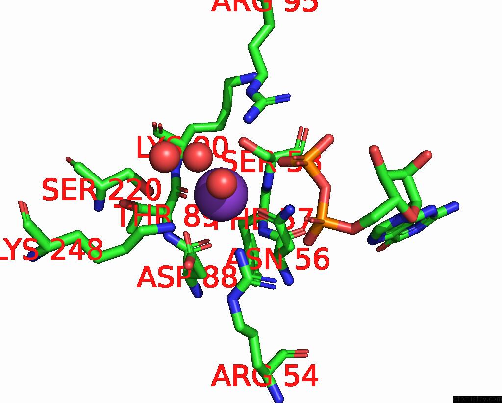
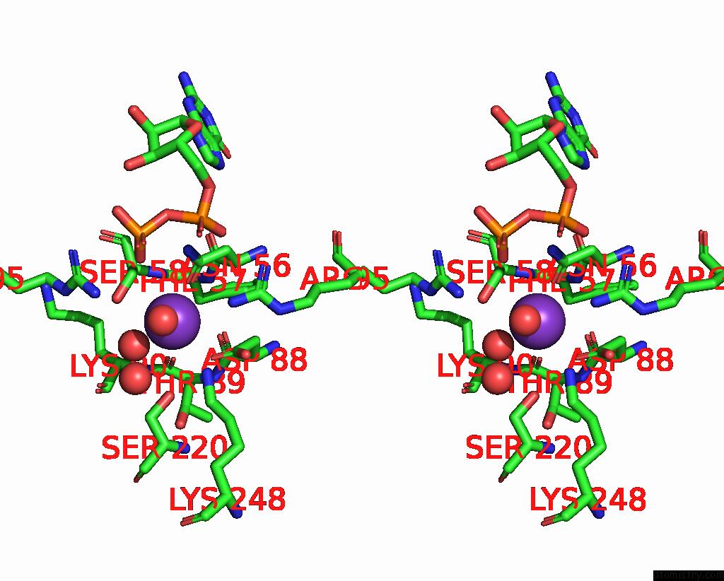
Potassium binding site 1 out of 4 in 8xw8
Go back to
Potassium binding site 1 out
of 4 in the Crystal Structure of Streptococcus Pneumoniae Pyruvate Kinase in Complex with Oxalate and Fructose 1,6-Bisphosphate and Gdp
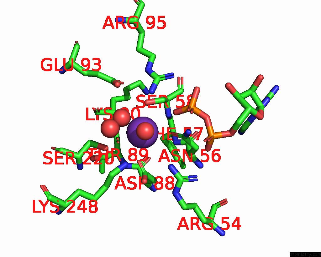
Mono view
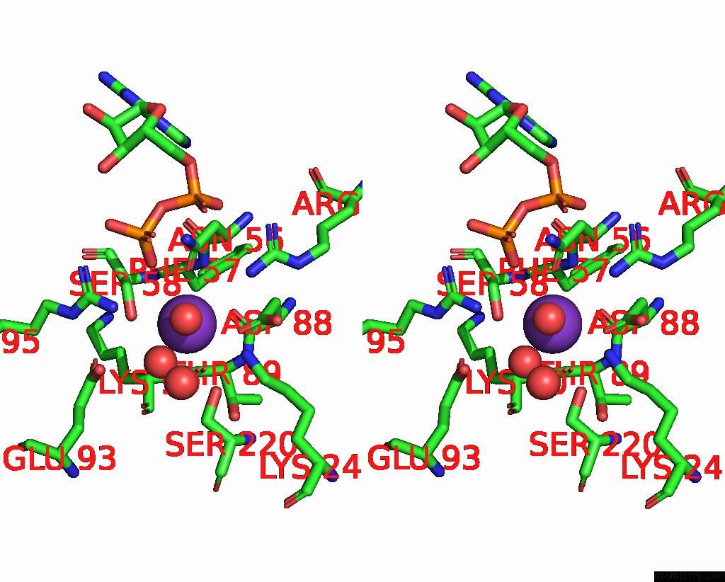
Stereo pair view
Mono view
Stereo pair view
A full contact list of Potassium with other atoms in the K binding
site number 1 of Crystal Structure of Streptococcus Pneumoniae Pyruvate Kinase in Complex with Oxalate and Fructose 1,6-Bisphosphate and Gdp within 5.0Å range:
|
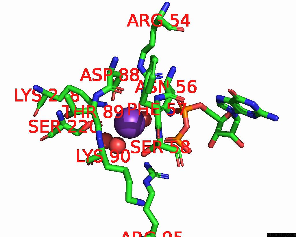
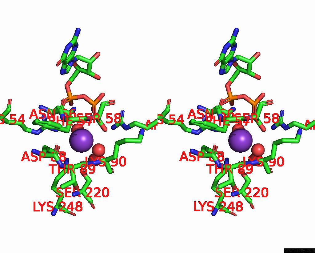
Potassium binding site 2 out of 4 in 8xw8
Go back to
Potassium binding site 2 out
of 4 in the Crystal Structure of Streptococcus Pneumoniae Pyruvate Kinase in Complex with Oxalate and Fructose 1,6-Bisphosphate and Gdp
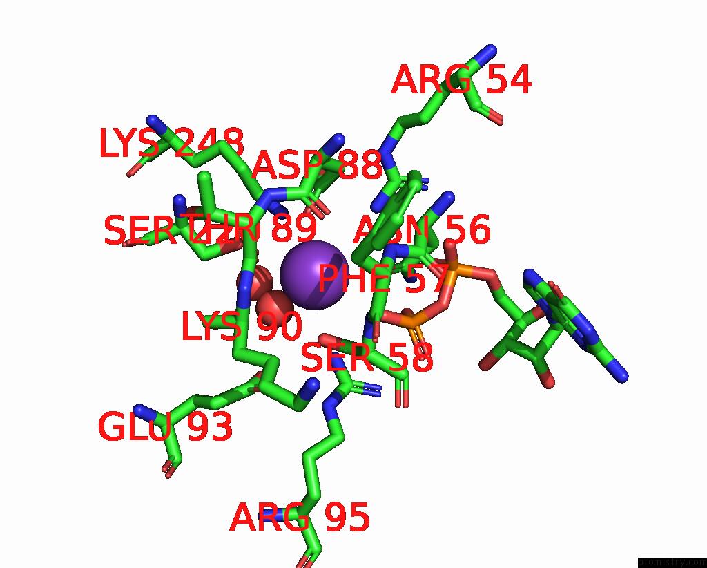
Mono view
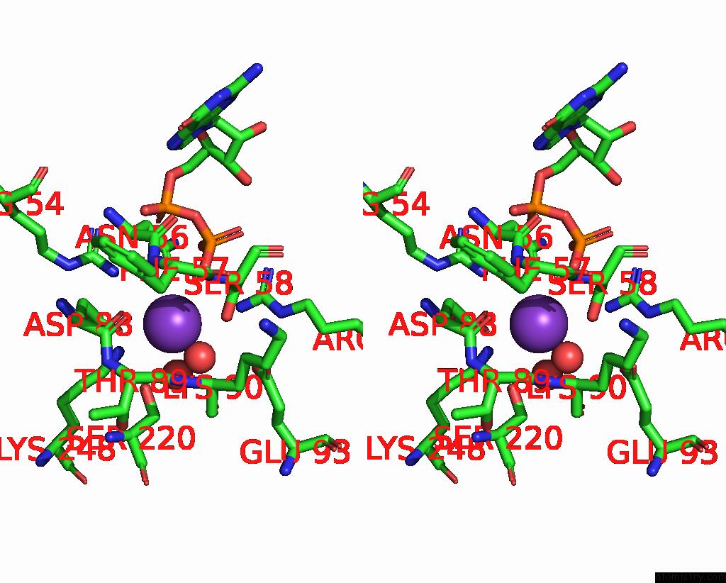
Stereo pair view
Mono view
Stereo pair view
A full contact list of Potassium with other atoms in the K binding
site number 2 of Crystal Structure of Streptococcus Pneumoniae Pyruvate Kinase in Complex with Oxalate and Fructose 1,6-Bisphosphate and Gdp within 5.0Å range:
|
Potassium binding site 3 out of 4 in 8xw8
Go back to
Potassium binding site 3 out
of 4 in the Crystal Structure of Streptococcus Pneumoniae Pyruvate Kinase in Complex with Oxalate and Fructose 1,6-Bisphosphate and Gdp
Mono view
Stereo pair view
Mono view
Stereo pair view
A full contact list of Potassium with other atoms in the K binding
site number 3 of Crystal Structure of Streptococcus Pneumoniae Pyruvate Kinase in Complex with Oxalate and Fructose 1,6-Bisphosphate and Gdp within 5.0Å range:
|
Potassium binding site 4 out of 4 in 8xw8
Go back to
Potassium binding site 4 out
of 4 in the Crystal Structure of Streptococcus Pneumoniae Pyruvate Kinase in Complex with Oxalate and Fructose 1,6-Bisphosphate and Gdp
Mono view
Stereo pair view
Mono view
Stereo pair view
A full contact list of Potassium with other atoms in the K binding
site number 4 of Crystal Structure of Streptococcus Pneumoniae Pyruvate Kinase in Complex with Oxalate and Fructose 1,6-Bisphosphate and Gdp within 5.0Å range:
|
Reference:
A.Taguchi,
R.Nakashima,
K.Nishino.
Structural Basis of Nucleotide Selectivity in Pyruvate Kinase. J.Mol.Biol. 68708 2024.
ISSN: ESSN 1089-8638
PubMed: 39009072
DOI: 10.1016/J.JMB.2024.168708
Page generated: Tue Aug 13 01:18:30 2024
ISSN: ESSN 1089-8638
PubMed: 39009072
DOI: 10.1016/J.JMB.2024.168708
Last articles
Zn in 9MJ5Zn in 9HNW
Zn in 9G0L
Zn in 9FNE
Zn in 9DZN
Zn in 9E0I
Zn in 9D32
Zn in 9DAK
Zn in 8ZXC
Zn in 8ZUF