Potassium in PDB 8olj: Crystal Structure of Archaeoglobus Fulgidus Afago-N Protein Representing N-L1-L2 Domains
Protein crystallography data
The structure of Crystal Structure of Archaeoglobus Fulgidus Afago-N Protein Representing N-L1-L2 Domains, PDB code: 8olj
was solved by
E.N.Manakova,
M.Zaremba,
S.Grazulis,
with X-Ray Crystallography technique. A brief refinement statistics is given in the table below:
| Resolution Low / High (Å) | 53.70 / 1.40 |
| Space group | P 32 2 1 |
| Cell size a, b, c (Å), α, β, γ (°) | 75.264, 75.264, 94.722, 90, 90, 120 |
| R / Rfree (%) | 14.5 / 17.8 |
Potassium Binding Sites:
The binding sites of Potassium atom in the Crystal Structure of Archaeoglobus Fulgidus Afago-N Protein Representing N-L1-L2 Domains
(pdb code 8olj). This binding sites where shown within
5.0 Angstroms radius around Potassium atom.
In total 3 binding sites of Potassium where determined in the Crystal Structure of Archaeoglobus Fulgidus Afago-N Protein Representing N-L1-L2 Domains, PDB code: 8olj:
Jump to Potassium binding site number: 1; 2; 3;
In total 3 binding sites of Potassium where determined in the Crystal Structure of Archaeoglobus Fulgidus Afago-N Protein Representing N-L1-L2 Domains, PDB code: 8olj:
Jump to Potassium binding site number: 1; 2; 3;
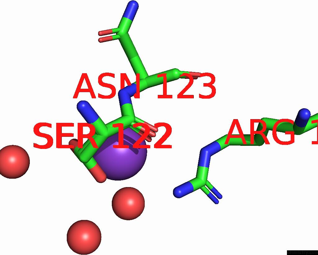
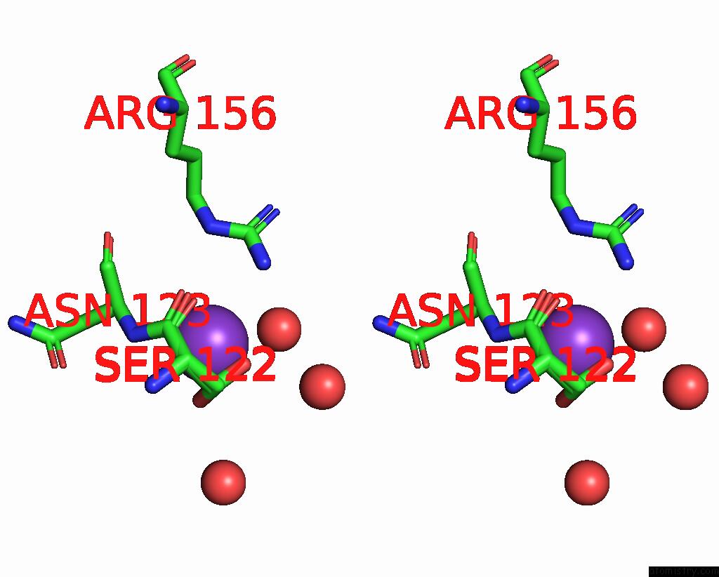
Potassium binding site 1 out of 3 in 8olj
Go back to
Potassium binding site 1 out
of 3 in the Crystal Structure of Archaeoglobus Fulgidus Afago-N Protein Representing N-L1-L2 Domains
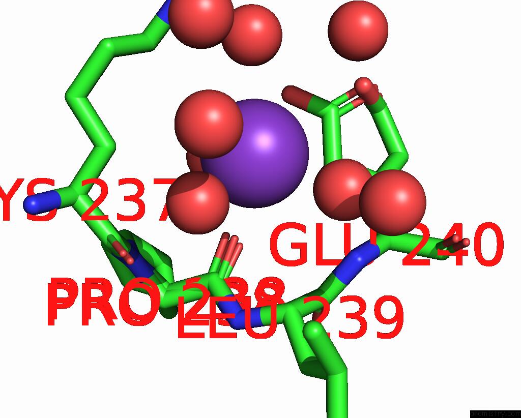
Mono view
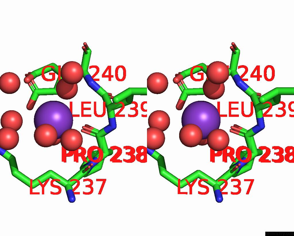
Stereo pair view
Mono view
Stereo pair view
A full contact list of Potassium with other atoms in the K binding
site number 1 of Crystal Structure of Archaeoglobus Fulgidus Afago-N Protein Representing N-L1-L2 Domains within 5.0Å range:
|
Potassium binding site 2 out of 3 in 8olj
Go back to
Potassium binding site 2 out
of 3 in the Crystal Structure of Archaeoglobus Fulgidus Afago-N Protein Representing N-L1-L2 Domains
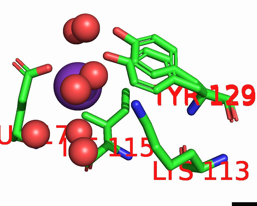
Mono view
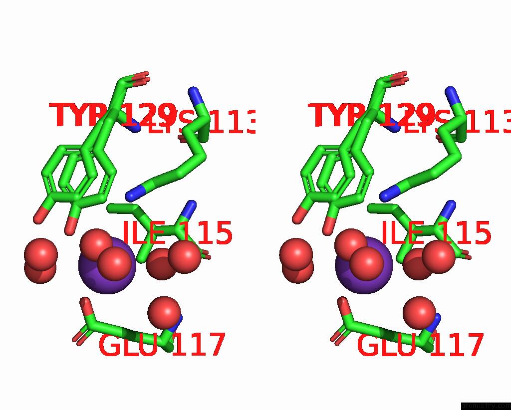
Stereo pair view
Mono view
Stereo pair view
A full contact list of Potassium with other atoms in the K binding
site number 2 of Crystal Structure of Archaeoglobus Fulgidus Afago-N Protein Representing N-L1-L2 Domains within 5.0Å range:
|
Potassium binding site 3 out of 3 in 8olj
Go back to
Potassium binding site 3 out
of 3 in the Crystal Structure of Archaeoglobus Fulgidus Afago-N Protein Representing N-L1-L2 Domains
Mono view
Stereo pair view
Mono view
Stereo pair view
A full contact list of Potassium with other atoms in the K binding
site number 3 of Crystal Structure of Archaeoglobus Fulgidus Afago-N Protein Representing N-L1-L2 Domains within 5.0Å range:
|
Reference:
E.Manakova,
E.Golovinas,
R.Poceviciute,
G.Sasnauskas,
A.Silanskas,
D.Rutkauskas,
M.Jankunec,
E.Zagorskaite,
E.Jurgelaitis,
A.Grybauskas,
C.Venclovas,
M.Zaremba.
The Missing Part: the Archaeoglobus Fulgidus Argonaute Forms A Functional Heterodimer with An N-L1-L2 Domain Protein. Nucleic Acids Res. 2024.
ISSN: ESSN 1362-4962
PubMed: 38197228
DOI: 10.1093/NAR/GKAD1241
Page generated: Tue Aug 13 00:14:03 2024
ISSN: ESSN 1362-4962
PubMed: 38197228
DOI: 10.1093/NAR/GKAD1241
Last articles
Zn in 9J0NZn in 9J0O
Zn in 9J0P
Zn in 9FJX
Zn in 9EKB
Zn in 9C0F
Zn in 9CAH
Zn in 9CH0
Zn in 9CH3
Zn in 9CH1