Potassium in PDB 8fxu: Thermoanaerobacter Thermosaccharolyticum Periplasmic Glucose-Binding Protein Glucose Complex: Badan Conjugate Attached at F17C
Protein crystallography data
The structure of Thermoanaerobacter Thermosaccharolyticum Periplasmic Glucose-Binding Protein Glucose Complex: Badan Conjugate Attached at F17C, PDB code: 8fxu
was solved by
M.J.Allert,
S.Kumar,
L.S.Beese,
H.W.Hellinga,
with X-Ray Crystallography technique. A brief refinement statistics is given in the table below:
| Resolution Low / High (Å) | 37.66 / 1.59 |
| Space group | P 21 21 21 |
| Cell size a, b, c (Å), α, β, γ (°) | 45.49, 53.06, 134.32, 90, 90, 90 |
| R / Rfree (%) | 16.1 / 17.8 |
Other elements in 8fxu:
The structure of Thermoanaerobacter Thermosaccharolyticum Periplasmic Glucose-Binding Protein Glucose Complex: Badan Conjugate Attached at F17C also contains other interesting chemical elements:
| Calcium | (Ca) | 1 atom |
| Chlorine | (Cl) | 2 atoms |
Potassium Binding Sites:
The binding sites of Potassium atom in the Thermoanaerobacter Thermosaccharolyticum Periplasmic Glucose-Binding Protein Glucose Complex: Badan Conjugate Attached at F17C
(pdb code 8fxu). This binding sites where shown within
5.0 Angstroms radius around Potassium atom.
In total only one binding site of Potassium was determined in the Thermoanaerobacter Thermosaccharolyticum Periplasmic Glucose-Binding Protein Glucose Complex: Badan Conjugate Attached at F17C, PDB code: 8fxu:
In total only one binding site of Potassium was determined in the Thermoanaerobacter Thermosaccharolyticum Periplasmic Glucose-Binding Protein Glucose Complex: Badan Conjugate Attached at F17C, PDB code: 8fxu:
Potassium binding site 1 out of 1 in 8fxu
Go back to
Potassium binding site 1 out
of 1 in the Thermoanaerobacter Thermosaccharolyticum Periplasmic Glucose-Binding Protein Glucose Complex: Badan Conjugate Attached at F17C
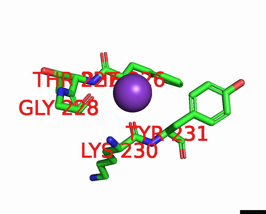
Mono view
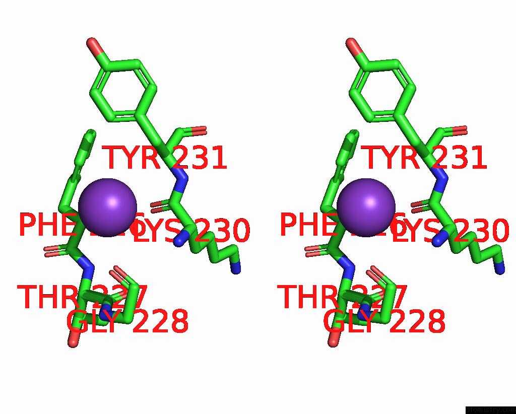
Stereo pair view
Mono view
Stereo pair view
A full contact list of Potassium with other atoms in the K binding
site number 1 of Thermoanaerobacter Thermosaccharolyticum Periplasmic Glucose-Binding Protein Glucose Complex: Badan Conjugate Attached at F17C within 5.0Å range:
|
Reference:
M.J.Allert,
S.Kumar,
Y.Wang,
L.S.Beese,
H.W.Hellinga.
Chromophore Carbonyl Twisting in Fluorescent Biosensors Encodes Direct Readout of Protein Conformations with Multicolor Switching. Commun Chem V. 6 168 2023.
ISSN: ESSN 2399-3669
PubMed: 37598249
DOI: 10.1038/S42004-023-00982-7
Page generated: Mon Aug 12 23:44:11 2024
ISSN: ESSN 2399-3669
PubMed: 37598249
DOI: 10.1038/S42004-023-00982-7
Last articles
Zn in 9MJ5Zn in 9HNW
Zn in 9G0L
Zn in 9FNE
Zn in 9DZN
Zn in 9E0I
Zn in 9D32
Zn in 9DAK
Zn in 8ZXC
Zn in 8ZUF