Potassium in PDB 8fsi: The Structure of A Crystallizable Variant of E. Coli Pyruvate Formate- Lyase Activating Enzyme Bound to Sam
Enzymatic activity of The Structure of A Crystallizable Variant of E. Coli Pyruvate Formate- Lyase Activating Enzyme Bound to Sam
All present enzymatic activity of The Structure of A Crystallizable Variant of E. Coli Pyruvate Formate- Lyase Activating Enzyme Bound to Sam:
1.97.1.4;
1.97.1.4;
Protein crystallography data
The structure of The Structure of A Crystallizable Variant of E. Coli Pyruvate Formate- Lyase Activating Enzyme Bound to Sam, PDB code: 8fsi
was solved by
J.D.Moody,
A.Galambas,
C.M.Lawrence,
J.B.Broderick,
with X-Ray Crystallography technique. A brief refinement statistics is given in the table below:
| Resolution Low / High (Å) | 41.77 / 1.46 |
| Space group | P 21 21 21 |
| Cell size a, b, c (Å), α, β, γ (°) | 46.405, 59.094, 83.545, 90, 90, 90 |
| R / Rfree (%) | 15.1 / 17.3 |
Other elements in 8fsi:
The structure of The Structure of A Crystallizable Variant of E. Coli Pyruvate Formate- Lyase Activating Enzyme Bound to Sam also contains other interesting chemical elements:
| Iron | (Fe) | 4 atoms |
| Chlorine | (Cl) | 1 atom |
Potassium Binding Sites:
The binding sites of Potassium atom in the The Structure of A Crystallizable Variant of E. Coli Pyruvate Formate- Lyase Activating Enzyme Bound to Sam
(pdb code 8fsi). This binding sites where shown within
5.0 Angstroms radius around Potassium atom.
In total 2 binding sites of Potassium where determined in the The Structure of A Crystallizable Variant of E. Coli Pyruvate Formate- Lyase Activating Enzyme Bound to Sam, PDB code: 8fsi:
Jump to Potassium binding site number: 1; 2;
In total 2 binding sites of Potassium where determined in the The Structure of A Crystallizable Variant of E. Coli Pyruvate Formate- Lyase Activating Enzyme Bound to Sam, PDB code: 8fsi:
Jump to Potassium binding site number: 1; 2;
Potassium binding site 1 out of 2 in 8fsi
Go back to
Potassium binding site 1 out
of 2 in the The Structure of A Crystallizable Variant of E. Coli Pyruvate Formate- Lyase Activating Enzyme Bound to Sam
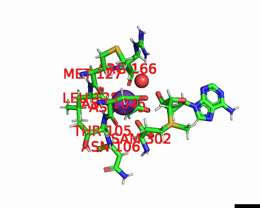
Mono view

Stereo pair view
Mono view
Stereo pair view
A full contact list of Potassium with other atoms in the K binding
site number 1 of The Structure of A Crystallizable Variant of E. Coli Pyruvate Formate- Lyase Activating Enzyme Bound to Sam within 5.0Å range:
|
Potassium binding site 2 out of 2 in 8fsi
Go back to
Potassium binding site 2 out
of 2 in the The Structure of A Crystallizable Variant of E. Coli Pyruvate Formate- Lyase Activating Enzyme Bound to Sam
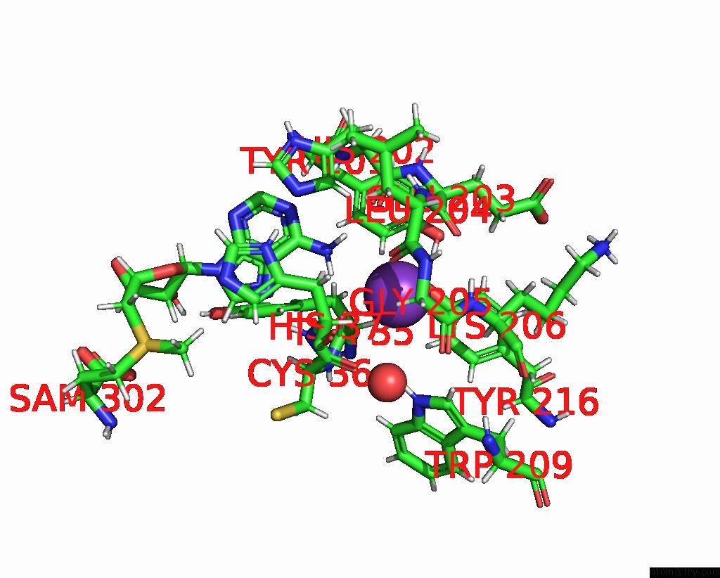
Mono view
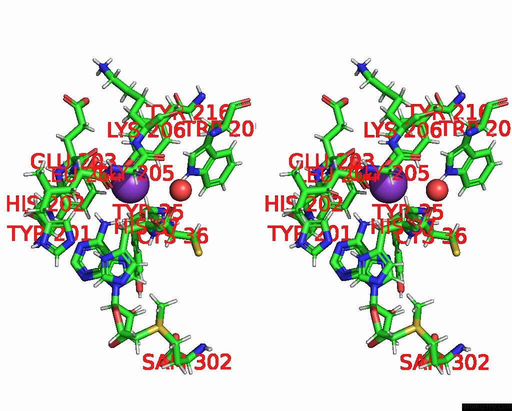
Stereo pair view
Mono view
Stereo pair view
A full contact list of Potassium with other atoms in the K binding
site number 2 of The Structure of A Crystallizable Variant of E. Coli Pyruvate Formate- Lyase Activating Enzyme Bound to Sam within 5.0Å range:
|
Reference:
J.D.Moody,
S.Hill,
M.N.Lundahl,
A.J.Saxton,
A.Galambas,
W.E.Broderick,
C.M.Lawrence,
J.B.Broderick.
Computational Engineering of Previously Crystallized Pyruvate Formate-Lyase Activating Enzyme Reveals Insights Into Sam Binding and Reductive Cleavage. J.Biol.Chem. 04791 2023.
ISSN: ESSN 1083-351X
PubMed: 37156396
DOI: 10.1016/J.JBC.2023.104791
Page generated: Mon Aug 12 23:43:53 2024
ISSN: ESSN 1083-351X
PubMed: 37156396
DOI: 10.1016/J.JBC.2023.104791
Last articles
Zn in 9MJ5Zn in 9HNW
Zn in 9G0L
Zn in 9FNE
Zn in 9DZN
Zn in 9E0I
Zn in 9D32
Zn in 9DAK
Zn in 8ZXC
Zn in 8ZUF