Potassium in PDB 7u59: Crystal Structure of Danio Rerio Histone Deacetylase 10 in Complex with Piperidine-4-Hydroxamic Acid Inhibitor
Enzymatic activity of Crystal Structure of Danio Rerio Histone Deacetylase 10 in Complex with Piperidine-4-Hydroxamic Acid Inhibitor
All present enzymatic activity of Crystal Structure of Danio Rerio Histone Deacetylase 10 in Complex with Piperidine-4-Hydroxamic Acid Inhibitor:
3.5.1.48; 3.5.1.62;
3.5.1.48; 3.5.1.62;
Protein crystallography data
The structure of Crystal Structure of Danio Rerio Histone Deacetylase 10 in Complex with Piperidine-4-Hydroxamic Acid Inhibitor, PDB code: 7u59
was solved by
C.J.Herbst-Gervasoni,
D.W.Christianson,
with X-Ray Crystallography technique. A brief refinement statistics is given in the table below:
| Resolution Low / High (Å) | 60.94 / 2.18 |
| Space group | P 31 2 1 |
| Cell size a, b, c (Å), α, β, γ (°) | 80.849, 80.849, 247.467, 90, 90, 120 |
| R / Rfree (%) | 17.9 / 21 |
Other elements in 7u59:
The structure of Crystal Structure of Danio Rerio Histone Deacetylase 10 in Complex with Piperidine-4-Hydroxamic Acid Inhibitor also contains other interesting chemical elements:
| Zinc | (Zn) | 1 atom |
| Sodium | (Na) | 1 atom |
Potassium Binding Sites:
The binding sites of Potassium atom in the Crystal Structure of Danio Rerio Histone Deacetylase 10 in Complex with Piperidine-4-Hydroxamic Acid Inhibitor
(pdb code 7u59). This binding sites where shown within
5.0 Angstroms radius around Potassium atom.
In total 2 binding sites of Potassium where determined in the Crystal Structure of Danio Rerio Histone Deacetylase 10 in Complex with Piperidine-4-Hydroxamic Acid Inhibitor, PDB code: 7u59:
Jump to Potassium binding site number: 1; 2;
In total 2 binding sites of Potassium where determined in the Crystal Structure of Danio Rerio Histone Deacetylase 10 in Complex with Piperidine-4-Hydroxamic Acid Inhibitor, PDB code: 7u59:
Jump to Potassium binding site number: 1; 2;
Potassium binding site 1 out of 2 in 7u59
Go back to
Potassium binding site 1 out
of 2 in the Crystal Structure of Danio Rerio Histone Deacetylase 10 in Complex with Piperidine-4-Hydroxamic Acid Inhibitor
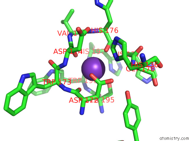
Mono view
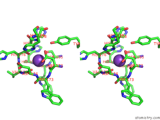
Stereo pair view
Mono view
Stereo pair view
A full contact list of Potassium with other atoms in the K binding
site number 1 of Crystal Structure of Danio Rerio Histone Deacetylase 10 in Complex with Piperidine-4-Hydroxamic Acid Inhibitor within 5.0Å range:
|
Potassium binding site 2 out of 2 in 7u59
Go back to
Potassium binding site 2 out
of 2 in the Crystal Structure of Danio Rerio Histone Deacetylase 10 in Complex with Piperidine-4-Hydroxamic Acid Inhibitor
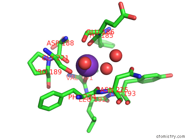
Mono view
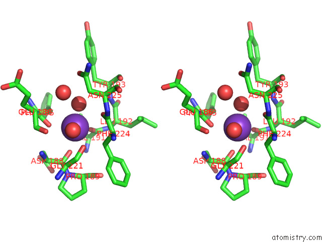
Stereo pair view
Mono view
Stereo pair view
A full contact list of Potassium with other atoms in the K binding
site number 2 of Crystal Structure of Danio Rerio Histone Deacetylase 10 in Complex with Piperidine-4-Hydroxamic Acid Inhibitor within 5.0Å range:
|
Reference:
D.Herp,
J.Ridinger,
D.Robaa,
S.A.Shinsky,
K.Schmidtkunz,
T.Z.Yesiloglu,
T.Bayer,
R.R.Steimbach,
C.J.Herbst-Gervasoni,
A.Merz,
C.Romier,
P.Sehr,
N.Gunkel,
A.K.Miller,
D.W.Christianson,
I.Oehme,
W.Sippl,
M.Jung.
First Fluorescent Acetylspermidine Deacetylation Assay For HDAC10 Identifies Selective Inhibitors with Cellular Target Engagement. Chembiochem V. 23 00180 2022.
ISSN: ESSN 1439-7633
PubMed: 35608330
DOI: 10.1002/CBIC.202200180
Page generated: Mon Aug 12 21:19:06 2024
ISSN: ESSN 1439-7633
PubMed: 35608330
DOI: 10.1002/CBIC.202200180
Last articles
Zn in 9MJ5Zn in 9HNW
Zn in 9G0L
Zn in 9FNE
Zn in 9DZN
Zn in 9E0I
Zn in 9D32
Zn in 9DAK
Zn in 8ZXC
Zn in 8ZUF