Potassium in PDB 7t7k: Structure of SPAC806.04C Protein From Fission Yeast Bound to CO2+
Protein crystallography data
The structure of Structure of SPAC806.04C Protein From Fission Yeast Bound to CO2+, PDB code: 7t7k
was solved by
A.Jacewicz,
A.M.Sanchez,
S.Shuman,
with X-Ray Crystallography technique. A brief refinement statistics is given in the table below:
| Resolution Low / High (Å) | 46.42 / 1.90 |
| Space group | P 2 21 21 |
| Cell size a, b, c (Å), α, β, γ (°) | 58.654, 116.314, 151.862, 90, 90, 90 |
| R / Rfree (%) | 16.4 / 19.6 |
Other elements in 7t7k:
The structure of Structure of SPAC806.04C Protein From Fission Yeast Bound to CO2+ also contains other interesting chemical elements:
| Cobalt | (Co) | 4 atoms |
| Chlorine | (Cl) | 1 atom |
Potassium Binding Sites:
The binding sites of Potassium atom in the Structure of SPAC806.04C Protein From Fission Yeast Bound to CO2+
(pdb code 7t7k). This binding sites where shown within
5.0 Angstroms radius around Potassium atom.
In total 4 binding sites of Potassium where determined in the Structure of SPAC806.04C Protein From Fission Yeast Bound to CO2+, PDB code: 7t7k:
Jump to Potassium binding site number: 1; 2; 3; 4;
In total 4 binding sites of Potassium where determined in the Structure of SPAC806.04C Protein From Fission Yeast Bound to CO2+, PDB code: 7t7k:
Jump to Potassium binding site number: 1; 2; 3; 4;
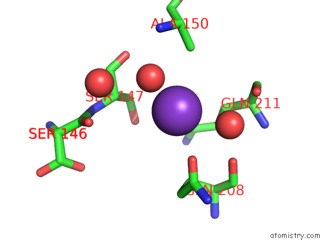
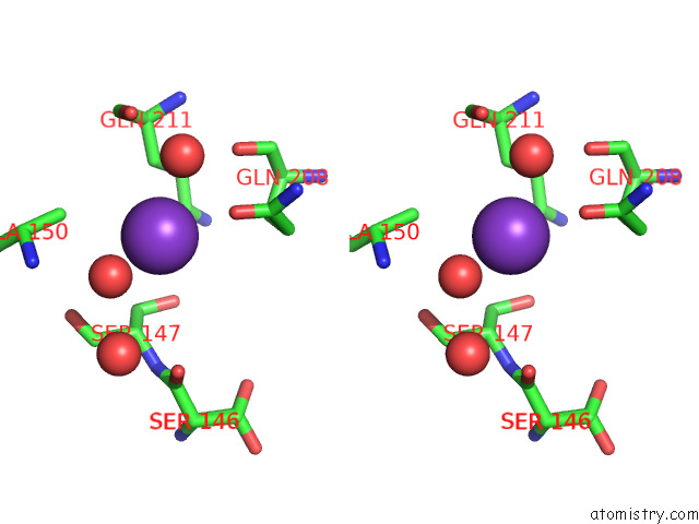
Potassium binding site 1 out of 4 in 7t7k
Go back to
Potassium binding site 1 out
of 4 in the Structure of SPAC806.04C Protein From Fission Yeast Bound to CO2+
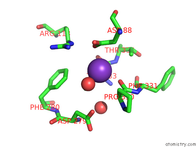
Mono view
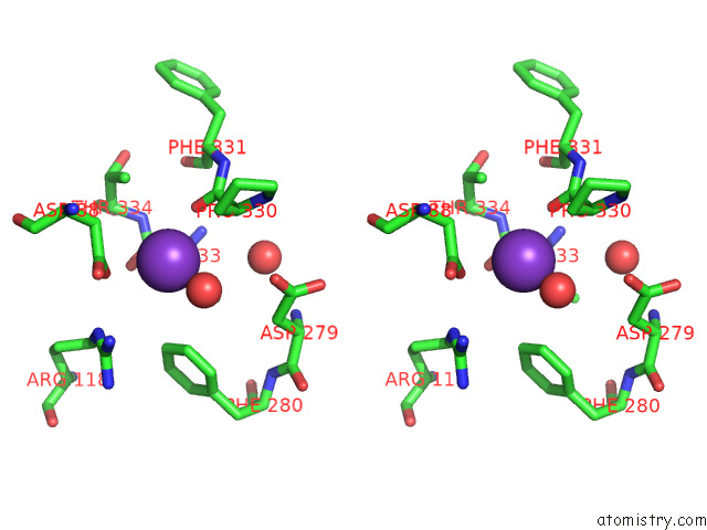
Stereo pair view
Mono view
Stereo pair view
A full contact list of Potassium with other atoms in the K binding
site number 1 of Structure of SPAC806.04C Protein From Fission Yeast Bound to CO2+ within 5.0Å range:
|
Potassium binding site 2 out of 4 in 7t7k
Go back to
Potassium binding site 2 out
of 4 in the Structure of SPAC806.04C Protein From Fission Yeast Bound to CO2+
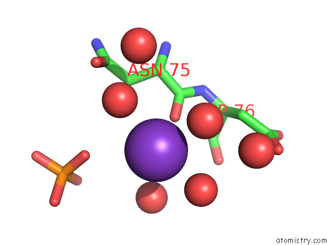
Mono view
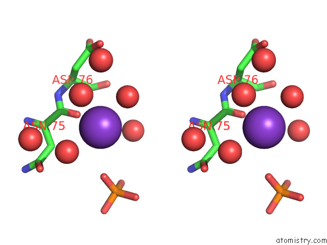
Stereo pair view
Mono view
Stereo pair view
A full contact list of Potassium with other atoms in the K binding
site number 2 of Structure of SPAC806.04C Protein From Fission Yeast Bound to CO2+ within 5.0Å range:
|
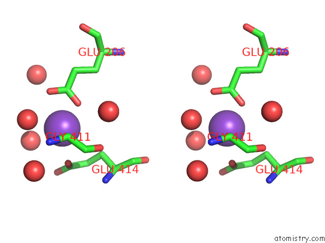
Potassium binding site 3 out of 4 in 7t7k
Go back to
Potassium binding site 3 out
of 4 in the Structure of SPAC806.04C Protein From Fission Yeast Bound to CO2+
Mono view
Stereo pair view
Mono view
Stereo pair view
A full contact list of Potassium with other atoms in the K binding
site number 3 of Structure of SPAC806.04C Protein From Fission Yeast Bound to CO2+ within 5.0Å range:
|
Potassium binding site 4 out of 4 in 7t7k
Go back to
Potassium binding site 4 out
of 4 in the Structure of SPAC806.04C Protein From Fission Yeast Bound to CO2+

Mono view
Stereo pair view
Mono view
Stereo pair view
A full contact list of Potassium with other atoms in the K binding
site number 4 of Structure of SPAC806.04C Protein From Fission Yeast Bound to CO2+ within 5.0Å range:
|
Reference:
A.M.Sanchez,
A.Jacewicz,
S.Shuman.
Fission Yeast DUF89 and DUF8901 Are Cobalt/Nickel-Dependent Phosphatase-Pyrophosphatases That Act Via A Covalent Aspartyl-Phosphate Intermediate. J.Biol.Chem. V. 298 01851 2022.
ISSN: ESSN 1083-351X
PubMed: 35314193
DOI: 10.1016/J.JBC.2022.101851
Page generated: Mon Aug 12 21:14:06 2024
ISSN: ESSN 1083-351X
PubMed: 35314193
DOI: 10.1016/J.JBC.2022.101851
Last articles
Zn in 9J0NZn in 9J0O
Zn in 9J0P
Zn in 9FJX
Zn in 9EKB
Zn in 9C0F
Zn in 9CAH
Zn in 9CH0
Zn in 9CH3
Zn in 9CH1