Potassium in PDB 7s10: Crystal Structure of Ascorbate Peroxidase Triple Mutant: S160M, L203M, Q204M
Enzymatic activity of Crystal Structure of Ascorbate Peroxidase Triple Mutant: S160M, L203M, Q204M
All present enzymatic activity of Crystal Structure of Ascorbate Peroxidase Triple Mutant: S160M, L203M, Q204M:
1.11.1.11;
1.11.1.11;
Protein crystallography data
The structure of Crystal Structure of Ascorbate Peroxidase Triple Mutant: S160M, L203M, Q204M, PDB code: 7s10
was solved by
T.L.Poulos,
J.Kim,
V.Murarka,
with X-Ray Crystallography technique. A brief refinement statistics is given in the table below:
| Resolution Low / High (Å) | 36.23 / 1.40 |
| Space group | P 42 21 2 |
| Cell size a, b, c (Å), α, β, γ (°) | 82.644, 82.644, 75.322, 90, 90, 90 |
| R / Rfree (%) | 17.1 / 20.8 |
Other elements in 7s10:
The structure of Crystal Structure of Ascorbate Peroxidase Triple Mutant: S160M, L203M, Q204M also contains other interesting chemical elements:
| Iron | (Fe) | 1 atom |
Potassium Binding Sites:
The binding sites of Potassium atom in the Crystal Structure of Ascorbate Peroxidase Triple Mutant: S160M, L203M, Q204M
(pdb code 7s10). This binding sites where shown within
5.0 Angstroms radius around Potassium atom.
In total only one binding site of Potassium was determined in the Crystal Structure of Ascorbate Peroxidase Triple Mutant: S160M, L203M, Q204M, PDB code: 7s10:
In total only one binding site of Potassium was determined in the Crystal Structure of Ascorbate Peroxidase Triple Mutant: S160M, L203M, Q204M, PDB code: 7s10:
Potassium binding site 1 out of 1 in 7s10
Go back to
Potassium binding site 1 out
of 1 in the Crystal Structure of Ascorbate Peroxidase Triple Mutant: S160M, L203M, Q204M
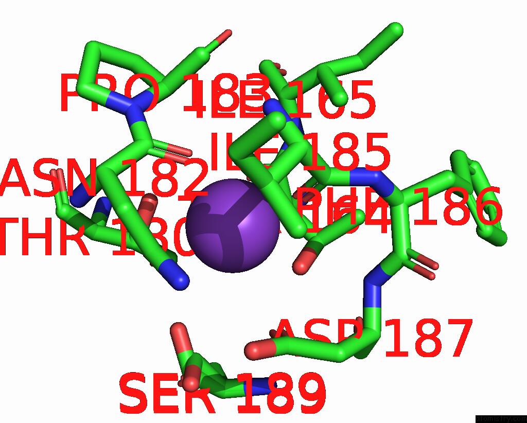
Mono view
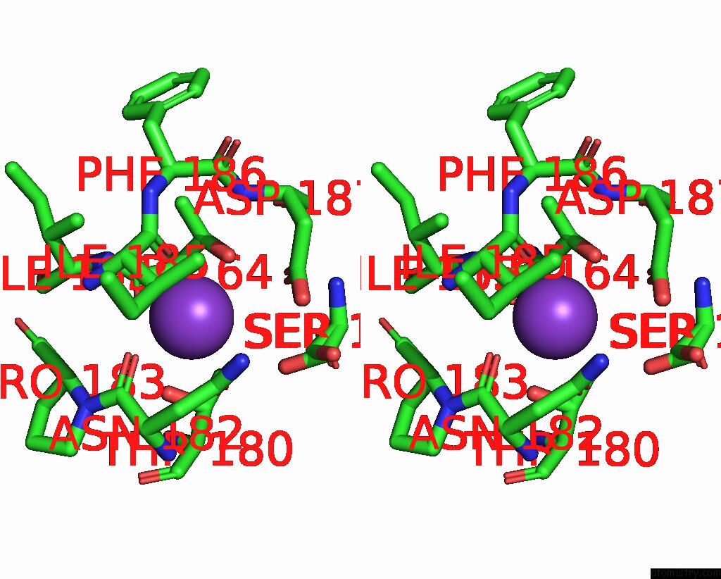
Stereo pair view
Mono view
Stereo pair view
A full contact list of Potassium with other atoms in the K binding
site number 1 of Crystal Structure of Ascorbate Peroxidase Triple Mutant: S160M, L203M, Q204M within 5.0Å range:
|
Reference:
T.L.Poulos,
J.S.Kim,
V.C.Murarka.
Computational Analysis of the Tryptophan Cation Radical Energetics in Peroxidase Compound I. J.Biol.Inorg.Chem. V. 27 229 2022.
ISSN: ESSN 1432-1327
PubMed: 35064363
DOI: 10.1007/S00775-022-01925-8
Page generated: Mon Aug 12 20:57:50 2024
ISSN: ESSN 1432-1327
PubMed: 35064363
DOI: 10.1007/S00775-022-01925-8
Last articles
Zn in 9J0NZn in 9J0O
Zn in 9J0P
Zn in 9FJX
Zn in 9EKB
Zn in 9C0F
Zn in 9CAH
Zn in 9CH0
Zn in 9CH3
Zn in 9CH1