Potassium in PDB 7rxv: Human Methionine Adenosyltransferase 2A Bound to Methylthioadenosine, Malonate (Mla) and MGF3
Enzymatic activity of Human Methionine Adenosyltransferase 2A Bound to Methylthioadenosine, Malonate (Mla) and MGF3
All present enzymatic activity of Human Methionine Adenosyltransferase 2A Bound to Methylthioadenosine, Malonate (Mla) and MGF3:
2.5.1.6;
2.5.1.6;
Protein crystallography data
The structure of Human Methionine Adenosyltransferase 2A Bound to Methylthioadenosine, Malonate (Mla) and MGF3, PDB code: 7rxv
was solved by
E.Fedorov,
C.N.Niland,
V.L.Schramm,
A.Ghosh,
with X-Ray Crystallography technique. A brief refinement statistics is given in the table below:
| Resolution Low / High (Å) | 19.86 / 1.07 |
| Space group | I 2 2 2 |
| Cell size a, b, c (Å), α, β, γ (°) | 67.981, 94.122, 117.315, 90, 90, 90 |
| R / Rfree (%) | 13 / 13.9 |
Other elements in 7rxv:
The structure of Human Methionine Adenosyltransferase 2A Bound to Methylthioadenosine, Malonate (Mla) and MGF3 also contains other interesting chemical elements:
| Fluorine | (F) | 3 atoms |
| Sodium | (Na) | 2 atoms |
| Magnesium | (Mg) | 2 atoms |
Potassium Binding Sites:
The binding sites of Potassium atom in the Human Methionine Adenosyltransferase 2A Bound to Methylthioadenosine, Malonate (Mla) and MGF3
(pdb code 7rxv). This binding sites where shown within
5.0 Angstroms radius around Potassium atom.
In total only one binding site of Potassium was determined in the Human Methionine Adenosyltransferase 2A Bound to Methylthioadenosine, Malonate (Mla) and MGF3, PDB code: 7rxv:
In total only one binding site of Potassium was determined in the Human Methionine Adenosyltransferase 2A Bound to Methylthioadenosine, Malonate (Mla) and MGF3, PDB code: 7rxv:
Potassium binding site 1 out of 1 in 7rxv
Go back to
Potassium binding site 1 out
of 1 in the Human Methionine Adenosyltransferase 2A Bound to Methylthioadenosine, Malonate (Mla) and MGF3
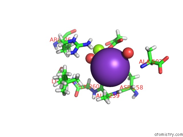
Mono view
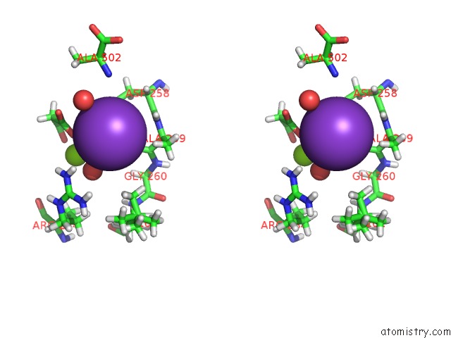
Stereo pair view
Mono view
Stereo pair view
A full contact list of Potassium with other atoms in the K binding
site number 1 of Human Methionine Adenosyltransferase 2A Bound to Methylthioadenosine, Malonate (Mla) and MGF3 within 5.0Å range:
|
Reference:
A.Ghosh,
C.N.Niland,
S.M.Cahill,
N.M.Karadkhelkar,
V.L.Schramm.
Mechanism of Triphosphate Hydrolysis By Human MAT2A at 1.07 Angstrom Resolution. J.Am.Chem.Soc. 2021.
ISSN: ESSN 1520-5126
PubMed: 34668717
DOI: 10.1021/JACS.1C09328
Page generated: Mon Aug 12 20:57:33 2024
ISSN: ESSN 1520-5126
PubMed: 34668717
DOI: 10.1021/JACS.1C09328
Last articles
Zn in 9J0NZn in 9J0O
Zn in 9J0P
Zn in 9FJX
Zn in 9EKB
Zn in 9C0F
Zn in 9CAH
Zn in 9CH0
Zn in 9CH3
Zn in 9CH1