Potassium in PDB 7q1b: Crystal Structure of Trypanosoma Cruzi Histone Deacetylase DAC2 Complexed with Quisinostat
Protein crystallography data
The structure of Crystal Structure of Trypanosoma Cruzi Histone Deacetylase DAC2 Complexed with Quisinostat, PDB code: 7q1b
was solved by
M.Marek,
E.Ramos-Morales,
C.Romier,
with X-Ray Crystallography technique. A brief refinement statistics is given in the table below:
| Resolution Low / High (Å) | 46.79 / 1.75 |
| Space group | I 2 2 2 |
| Cell size a, b, c (Å), α, β, γ (°) | 82.167, 93.584, 119.462, 90, 90, 90 |
| R / Rfree (%) | 19.2 / 21.8 |
Other elements in 7q1b:
The structure of Crystal Structure of Trypanosoma Cruzi Histone Deacetylase DAC2 Complexed with Quisinostat also contains other interesting chemical elements:
| Zinc | (Zn) | 1 atom |
Potassium Binding Sites:
The binding sites of Potassium atom in the Crystal Structure of Trypanosoma Cruzi Histone Deacetylase DAC2 Complexed with Quisinostat
(pdb code 7q1b). This binding sites where shown within
5.0 Angstroms radius around Potassium atom.
In total 2 binding sites of Potassium where determined in the Crystal Structure of Trypanosoma Cruzi Histone Deacetylase DAC2 Complexed with Quisinostat, PDB code: 7q1b:
Jump to Potassium binding site number: 1; 2;
In total 2 binding sites of Potassium where determined in the Crystal Structure of Trypanosoma Cruzi Histone Deacetylase DAC2 Complexed with Quisinostat, PDB code: 7q1b:
Jump to Potassium binding site number: 1; 2;
Potassium binding site 1 out of 2 in 7q1b
Go back to
Potassium binding site 1 out
of 2 in the Crystal Structure of Trypanosoma Cruzi Histone Deacetylase DAC2 Complexed with Quisinostat
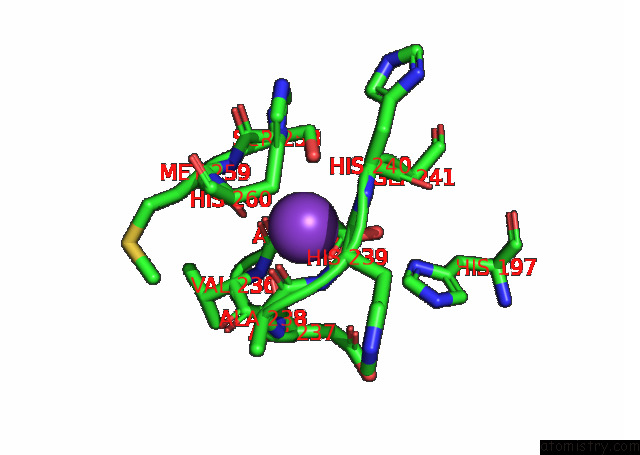
Mono view
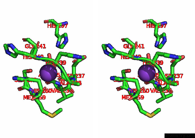
Stereo pair view
Mono view
Stereo pair view
A full contact list of Potassium with other atoms in the K binding
site number 1 of Crystal Structure of Trypanosoma Cruzi Histone Deacetylase DAC2 Complexed with Quisinostat within 5.0Å range:
|
Potassium binding site 2 out of 2 in 7q1b
Go back to
Potassium binding site 2 out
of 2 in the Crystal Structure of Trypanosoma Cruzi Histone Deacetylase DAC2 Complexed with Quisinostat
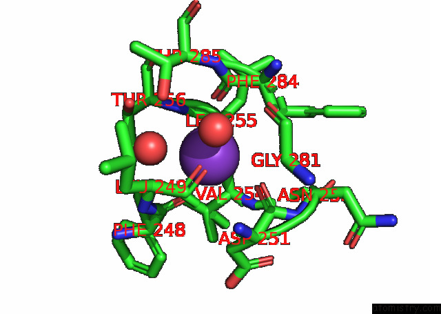
Mono view
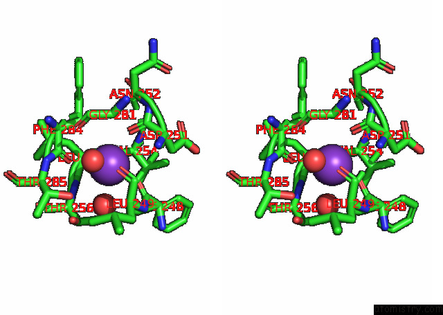
Stereo pair view
Mono view
Stereo pair view
A full contact list of Potassium with other atoms in the K binding
site number 2 of Crystal Structure of Trypanosoma Cruzi Histone Deacetylase DAC2 Complexed with Quisinostat within 5.0Å range:
|
Reference:
M.Marek,
E.Ramos-Morales,
C.Romier.
Species-Selective Targeting of Pathogens Revealed By the Atypical Structure and Active Site of Trypanosoma Cruzi Histone Deacetylase DAC2 To Be Published.
Page generated: Mon Aug 12 20:26:21 2024
Last articles
Zn in 9MJ5Zn in 9HNW
Zn in 9G0L
Zn in 9FNE
Zn in 9DZN
Zn in 9E0I
Zn in 9D32
Zn in 9DAK
Zn in 8ZXC
Zn in 8ZUF