Potassium in PDB 7q0g: Crystal Structure of the Receptor Binding Domain of Sars-Cov-2 Beta Variant Spike Glycoprotein in Complex with Beta-49 and Fi-3A Fabs
Protein crystallography data
The structure of Crystal Structure of the Receptor Binding Domain of Sars-Cov-2 Beta Variant Spike Glycoprotein in Complex with Beta-49 and Fi-3A Fabs, PDB code: 7q0g
was solved by
D.Zhou,
J.Ren,
D.I.Stuart,
with X-Ray Crystallography technique. A brief refinement statistics is given in the table below:
| Resolution Low / High (Å) | 107.81 / 1.82 |
| Space group | P 21 21 21 |
| Cell size a, b, c (Å), α, β, γ (°) | 89.292, 106.538, 215.611, 90, 90, 90 |
| R / Rfree (%) | 19.2 / 22.4 |
Other elements in 7q0g:
The structure of Crystal Structure of the Receptor Binding Domain of Sars-Cov-2 Beta Variant Spike Glycoprotein in Complex with Beta-49 and Fi-3A Fabs also contains other interesting chemical elements:
| Chlorine | (Cl) | 14 atoms |
Potassium Binding Sites:
Pages:
>>> Page 1 <<< Page 2, Binding sites: 11 - 14;Binding sites:
The binding sites of Potassium atom in the Crystal Structure of the Receptor Binding Domain of Sars-Cov-2 Beta Variant Spike Glycoprotein in Complex with Beta-49 and Fi-3A Fabs (pdb code 7q0g). This binding sites where shown within 5.0 Angstroms radius around Potassium atom.In total 14 binding sites of Potassium where determined in the Crystal Structure of the Receptor Binding Domain of Sars-Cov-2 Beta Variant Spike Glycoprotein in Complex with Beta-49 and Fi-3A Fabs, PDB code: 7q0g:
Jump to Potassium binding site number: 1; 2; 3; 4; 5; 6; 7; 8; 9; 10;
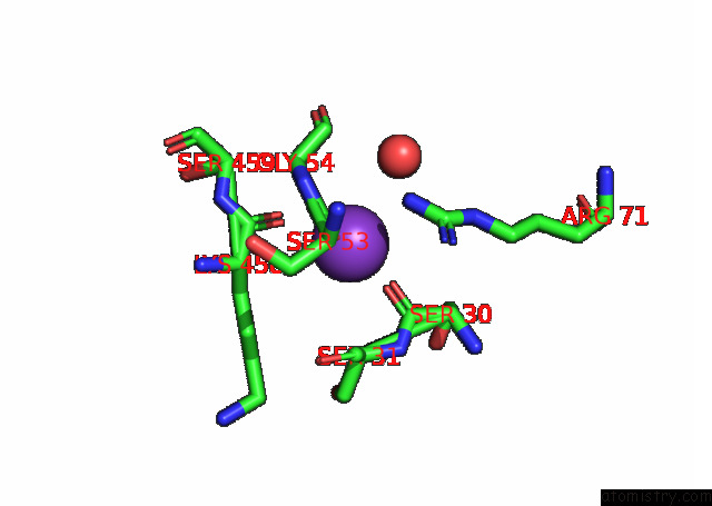
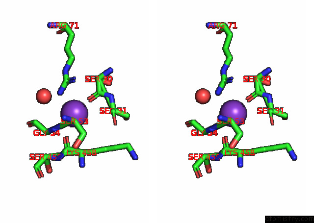
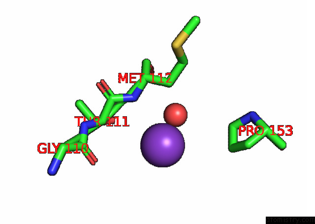
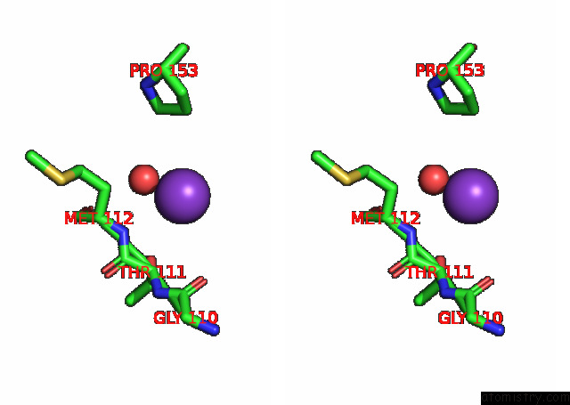
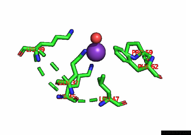
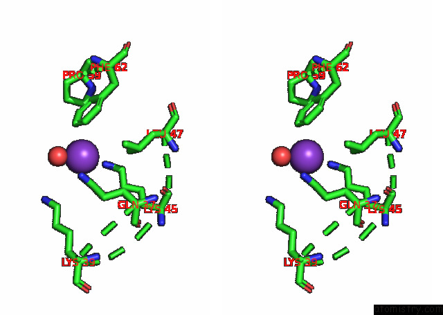
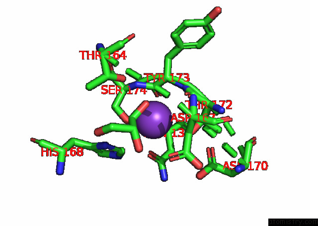
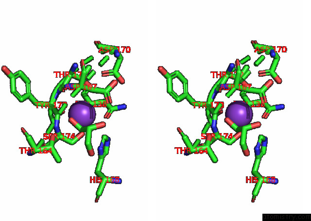
Potassium binding site 1 out of 14 in 7q0g
Go back to
Potassium binding site 1 out
of 14 in the Crystal Structure of the Receptor Binding Domain of Sars-Cov-2 Beta Variant Spike Glycoprotein in Complex with Beta-49 and Fi-3A Fabs
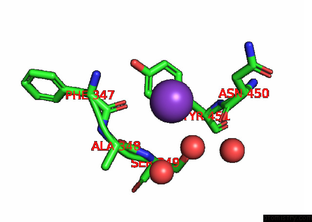
Mono view
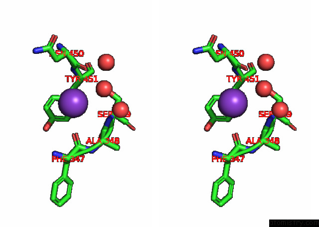
Stereo pair view
Mono view
Stereo pair view
A full contact list of Potassium with other atoms in the K binding
site number 1 of Crystal Structure of the Receptor Binding Domain of Sars-Cov-2 Beta Variant Spike Glycoprotein in Complex with Beta-49 and Fi-3A Fabs within 5.0Å range:
|
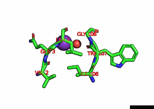
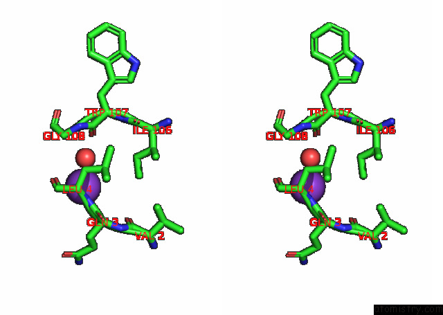
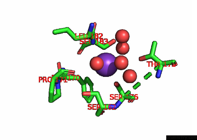
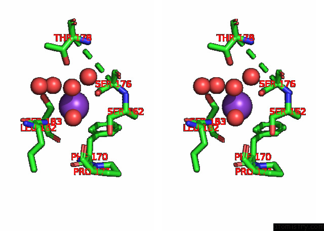
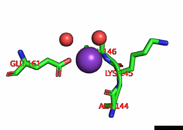
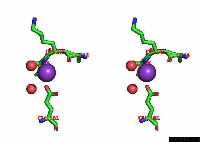
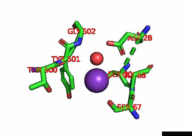
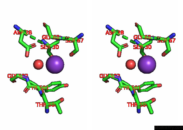
Potassium binding site 2 out of 14 in 7q0g
Go back to
Potassium binding site 2 out
of 14 in the Crystal Structure of the Receptor Binding Domain of Sars-Cov-2 Beta Variant Spike Glycoprotein in Complex with Beta-49 and Fi-3A Fabs
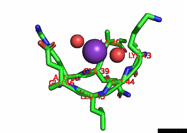
Mono view
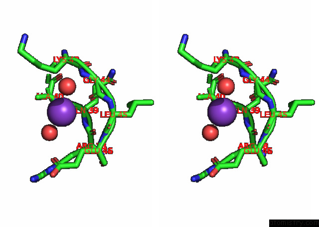
Stereo pair view
Mono view
Stereo pair view
A full contact list of Potassium with other atoms in the K binding
site number 2 of Crystal Structure of the Receptor Binding Domain of Sars-Cov-2 Beta Variant Spike Glycoprotein in Complex with Beta-49 and Fi-3A Fabs within 5.0Å range:
|
Potassium binding site 3 out of 14 in 7q0g
Go back to
Potassium binding site 3 out
of 14 in the Crystal Structure of the Receptor Binding Domain of Sars-Cov-2 Beta Variant Spike Glycoprotein in Complex with Beta-49 and Fi-3A Fabs
Mono view
Stereo pair view
Mono view
Stereo pair view
A full contact list of Potassium with other atoms in the K binding
site number 3 of Crystal Structure of the Receptor Binding Domain of Sars-Cov-2 Beta Variant Spike Glycoprotein in Complex with Beta-49 and Fi-3A Fabs within 5.0Å range:
|
Potassium binding site 4 out of 14 in 7q0g
Go back to
Potassium binding site 4 out
of 14 in the Crystal Structure of the Receptor Binding Domain of Sars-Cov-2 Beta Variant Spike Glycoprotein in Complex with Beta-49 and Fi-3A Fabs
Mono view
Stereo pair view
Mono view
Stereo pair view
A full contact list of Potassium with other atoms in the K binding
site number 4 of Crystal Structure of the Receptor Binding Domain of Sars-Cov-2 Beta Variant Spike Glycoprotein in Complex with Beta-49 and Fi-3A Fabs within 5.0Å range:
|
Potassium binding site 5 out of 14 in 7q0g
Go back to
Potassium binding site 5 out
of 14 in the Crystal Structure of the Receptor Binding Domain of Sars-Cov-2 Beta Variant Spike Glycoprotein in Complex with Beta-49 and Fi-3A Fabs
Mono view
Stereo pair view
Mono view
Stereo pair view
A full contact list of Potassium with other atoms in the K binding
site number 5 of Crystal Structure of the Receptor Binding Domain of Sars-Cov-2 Beta Variant Spike Glycoprotein in Complex with Beta-49 and Fi-3A Fabs within 5.0Å range:
|
Potassium binding site 6 out of 14 in 7q0g
Go back to
Potassium binding site 6 out
of 14 in the Crystal Structure of the Receptor Binding Domain of Sars-Cov-2 Beta Variant Spike Glycoprotein in Complex with Beta-49 and Fi-3A Fabs
Mono view
Stereo pair view
Mono view
Stereo pair view
A full contact list of Potassium with other atoms in the K binding
site number 6 of Crystal Structure of the Receptor Binding Domain of Sars-Cov-2 Beta Variant Spike Glycoprotein in Complex with Beta-49 and Fi-3A Fabs within 5.0Å range:
|
Potassium binding site 7 out of 14 in 7q0g
Go back to
Potassium binding site 7 out
of 14 in the Crystal Structure of the Receptor Binding Domain of Sars-Cov-2 Beta Variant Spike Glycoprotein in Complex with Beta-49 and Fi-3A Fabs
Mono view
Stereo pair view
Mono view
Stereo pair view
A full contact list of Potassium with other atoms in the K binding
site number 7 of Crystal Structure of the Receptor Binding Domain of Sars-Cov-2 Beta Variant Spike Glycoprotein in Complex with Beta-49 and Fi-3A Fabs within 5.0Å range:
|
Potassium binding site 8 out of 14 in 7q0g
Go back to
Potassium binding site 8 out
of 14 in the Crystal Structure of the Receptor Binding Domain of Sars-Cov-2 Beta Variant Spike Glycoprotein in Complex with Beta-49 and Fi-3A Fabs
Mono view
Stereo pair view
Mono view
Stereo pair view
A full contact list of Potassium with other atoms in the K binding
site number 8 of Crystal Structure of the Receptor Binding Domain of Sars-Cov-2 Beta Variant Spike Glycoprotein in Complex with Beta-49 and Fi-3A Fabs within 5.0Å range:
|
Potassium binding site 9 out of 14 in 7q0g
Go back to
Potassium binding site 9 out
of 14 in the Crystal Structure of the Receptor Binding Domain of Sars-Cov-2 Beta Variant Spike Glycoprotein in Complex with Beta-49 and Fi-3A Fabs
Mono view
Stereo pair view
Mono view
Stereo pair view
A full contact list of Potassium with other atoms in the K binding
site number 9 of Crystal Structure of the Receptor Binding Domain of Sars-Cov-2 Beta Variant Spike Glycoprotein in Complex with Beta-49 and Fi-3A Fabs within 5.0Å range:
|
Potassium binding site 10 out of 14 in 7q0g
Go back to
Potassium binding site 10 out
of 14 in the Crystal Structure of the Receptor Binding Domain of Sars-Cov-2 Beta Variant Spike Glycoprotein in Complex with Beta-49 and Fi-3A Fabs
Mono view
Stereo pair view
Mono view
Stereo pair view
A full contact list of Potassium with other atoms in the K binding
site number 10 of Crystal Structure of the Receptor Binding Domain of Sars-Cov-2 Beta Variant Spike Glycoprotein in Complex with Beta-49 and Fi-3A Fabs within 5.0Å range:
|
Reference:
C.Liu,
D.Zhou,
R.Nutalai,
H.M.Duyvesteyn,
A.Tuekprakhon,
H.M.Ginn,
W.Dejnirattisai,
P.Supasa,
A.J.Mentzer,
B.Wang,
J.B.Case,
Y.Zhao,
D.T.Skelly,
R.E.Chen,
S.A.Johnson,
T.G.Ritter,
C.Mason,
T.Malik,
N.Temperton,
N.G.Paterson,
M.A.Williams,
D.R.Hall,
D.K.Clare,
A.Howe,
P.J.Goulder,
E.E.Fry,
M.S.Diamond,
J.Mongkolsapaya,
J.Ren,
D.I.Stuart,
G.R.Screaton.
The Antibody Response to Sars-Cov-2 Beta Underscores the Antigenic Distance to Other Variants Cell Host Microbe 2021.
ISSN: ESSN 1934-6069
DOI: 10.1016/J.CHOM.2021.11.013
Page generated: Mon Aug 12 20:26:20 2024
ISSN: ESSN 1934-6069
DOI: 10.1016/J.CHOM.2021.11.013
Last articles
Zn in 9MJ5Zn in 9HNW
Zn in 9G0L
Zn in 9FNE
Zn in 9DZN
Zn in 9E0I
Zn in 9D32
Zn in 9DAK
Zn in 8ZXC
Zn in 8ZUF