Potassium in PDB 7jom: Crystal Structure of Danio Rerio Histone Deacetylase 6 Catalytic Domain 2 Complexed with to-317
Protein crystallography data
The structure of Crystal Structure of Danio Rerio Histone Deacetylase 6 Catalytic Domain 2 Complexed with to-317, PDB code: 7jom
was solved by
P.R.Watson,
D.W.Christianson,
with X-Ray Crystallography technique. A brief refinement statistics is given in the table below:
Resolution Low / High (Å)
|
59.43 /
1.84
|
Space group
|
P 21 21 21
|
Cell size a, b, c (Å), α, β, γ (°)
|
75.46,
96.45,
96.45,
90,
90,
90
|
R / Rfree (%)
|
16.8 /
20.7
|
Other elements in 7jom:
The structure of Crystal Structure of Danio Rerio Histone Deacetylase 6 Catalytic Domain 2 Complexed with to-317 also contains other interesting chemical elements:
Potassium Binding Sites:
The binding sites of Potassium atom in the Crystal Structure of Danio Rerio Histone Deacetylase 6 Catalytic Domain 2 Complexed with to-317
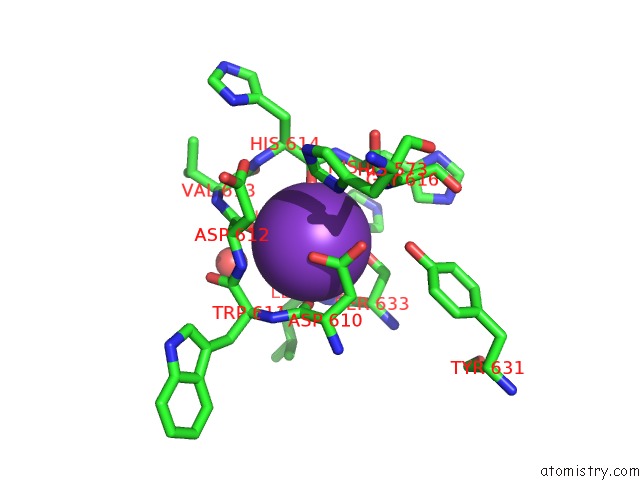
(pdb code 7jom). This binding sites where shown within
5.0 Angstroms radius around Potassium atom.
In total 5 binding sites of Potassium where determined in the
Crystal Structure of Danio Rerio Histone Deacetylase 6 Catalytic Domain 2 Complexed with to-317, PDB code: 7jom:
Jump to Potassium binding site number:
1;
2;
3;
4;
5;
Potassium binding site 1 out
of 5 in 7jom
Go back to
Potassium Binding Sites List in 7jom
Potassium binding site 1 out
of 5 in the Crystal Structure of Danio Rerio Histone Deacetylase 6 Catalytic Domain 2 Complexed with to-317
Mono view
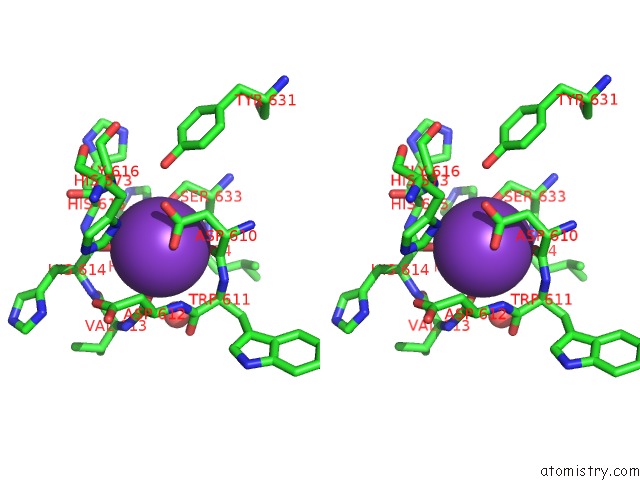
Stereo pair view
|
A full contact list of Potassium with other atoms in the K binding
site number 1 of Crystal Structure of Danio Rerio Histone Deacetylase 6 Catalytic Domain 2 Complexed with to-317 within 5.0Å range:
probe
|
atom
|
residue
|
distance (Å)
|
B
|
Occ
|
A:K908
b:6.8
occ:1.00
|
O
|
A:ASP612
|
2.6
|
4.8
|
1.0
|
O
|
A:HIS614
|
2.7
|
5.7
|
1.0
|
O
|
A:LEU634
|
2.7
|
7.6
|
1.0
|
OG
|
A:SER633
|
2.8
|
7.1
|
1.0
|
OD1
|
A:ASP610
|
2.8
|
6.4
|
1.0
|
O
|
A:ASP610
|
2.8
|
4.5
|
1.0
|
CG
|
A:ASP610
|
3.3
|
6.3
|
1.0
|
C
|
A:ASP610
|
3.5
|
5.4
|
1.0
|
C
|
A:ASP612
|
3.6
|
4.9
|
1.0
|
C
|
A:HIS614
|
3.7
|
5.7
|
1.0
|
C
|
A:LEU634
|
3.7
|
8.0
|
1.0
|
CB
|
A:ASP610
|
3.8
|
5.3
|
1.0
|
N
|
A:ASP612
|
3.8
|
4.6
|
1.0
|
OD2
|
A:ASP610
|
3.9
|
8.4
|
1.0
|
N
|
A:LEU634
|
3.9
|
6.5
|
1.0
|
CB
|
A:SER633
|
4.0
|
4.4
|
1.0
|
CB
|
A:HIS635
|
4.1
|
5.5
|
1.0
|
CA
|
A:ASP612
|
4.1
|
6.4
|
1.0
|
C
|
A:TRP611
|
4.2
|
5.0
|
1.0
|
N
|
A:TRP611
|
4.2
|
5.3
|
1.0
|
CB
|
A:ASP612
|
4.2
|
5.3
|
1.0
|
CA
|
A:ASP610
|
4.2
|
3.9
|
1.0
|
CA
|
A:HIS615
|
4.3
|
7.5
|
1.0
|
CA
|
A:TRP611
|
4.3
|
6.6
|
1.0
|
CA
|
A:SER633
|
4.3
|
5.5
|
1.0
|
N
|
A:HIS615
|
4.4
|
5.2
|
1.0
|
ND1
|
A:HIS635
|
4.4
|
9.4
|
1.0
|
N
|
A:GLY616
|
4.5
|
6.6
|
1.0
|
N
|
A:HIS614
|
4.5
|
4.7
|
1.0
|
O
|
A:HOH1046
|
4.5
|
7.2
|
1.0
|
CA
|
A:LEU634
|
4.5
|
7.5
|
1.0
|
C
|
A:SER633
|
4.5
|
7.6
|
1.0
|
CA
|
A:HIS635
|
4.5
|
6.3
|
1.0
|
N
|
A:HIS635
|
4.6
|
7.9
|
1.0
|
C
|
A:VAL613
|
4.7
|
5.5
|
1.0
|
CG
|
A:HIS635
|
4.7
|
7.0
|
1.0
|
N
|
A:VAL613
|
4.7
|
4.3
|
1.0
|
C
|
A:HIS615
|
4.7
|
7.0
|
1.0
|
OH
|
A:TYR631
|
4.7
|
6.2
|
1.0
|
CA
|
A:HIS614
|
4.7
|
4.8
|
1.0
|
O
|
A:TRP611
|
4.9
|
7.2
|
1.0
|
CE1
|
A:HIS573
|
4.9
|
4.9
|
1.0
|
|
Potassium binding site 2 out
of 5 in 7jom
Go back to
Potassium Binding Sites List in 7jom
Potassium binding site 2 out
of 5 in the Crystal Structure of Danio Rerio Histone Deacetylase 6 Catalytic Domain 2 Complexed with to-317
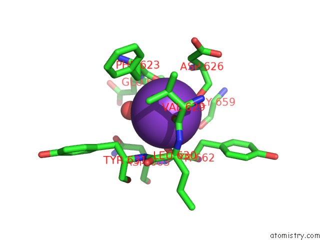
Mono view
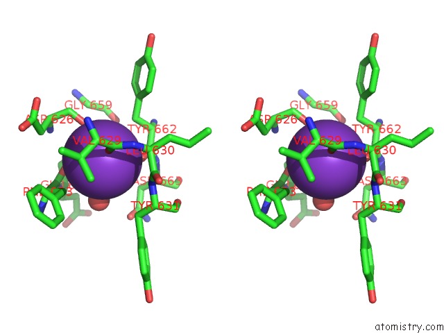
Stereo pair view
|
A full contact list of Potassium with other atoms in the K binding
site number 2 of Crystal Structure of Danio Rerio Histone Deacetylase 6 Catalytic Domain 2 Complexed with to-317 within 5.0Å range:
probe
|
atom
|
residue
|
distance (Å)
|
B
|
Occ
|
A:K909
b:7.5
occ:1.00
|
O
|
A:PHE623
|
2.7
|
8.1
|
1.0
|
O
|
A:VAL629
|
2.7
|
7.4
|
1.0
|
O
|
A:HOH1078
|
2.7
|
7.4
|
1.0
|
O
|
A:TYR662
|
2.8
|
6.3
|
1.0
|
O
|
A:ASP626
|
2.9
|
8.7
|
1.0
|
O
|
A:HOH1107
|
3.0
|
6.2
|
1.0
|
C
|
A:TYR662
|
3.6
|
8.2
|
1.0
|
C
|
A:PHE623
|
3.6
|
7.8
|
1.0
|
CB
|
A:TYR662
|
3.7
|
9.6
|
1.0
|
CB
|
A:PHE623
|
3.7
|
5.2
|
1.0
|
C
|
A:VAL629
|
3.9
|
5.5
|
1.0
|
C
|
A:ASP626
|
4.0
|
12.6
|
1.0
|
CA
|
A:TYR662
|
4.3
|
7.7
|
1.0
|
N
|
A:TYR631
|
4.3
|
4.4
|
1.0
|
CA
|
A:PHE623
|
4.3
|
7.1
|
1.0
|
N
|
A:ASP626
|
4.5
|
6.9
|
1.0
|
N
|
A:ASN663
|
4.5
|
7.0
|
1.0
|
N
|
A:GLU624
|
4.5
|
6.9
|
1.0
|
CA
|
A:GLU624
|
4.5
|
5.9
|
1.0
|
CA
|
A:ASP626
|
4.6
|
10.4
|
1.0
|
CA
|
A:LEU630
|
4.6
|
5.7
|
1.0
|
CB
|
A:ASP626
|
4.7
|
8.6
|
1.0
|
O
|
A:GLU624
|
4.7
|
8.9
|
1.0
|
C
|
A:GLU624
|
4.7
|
7.3
|
1.0
|
CB
|
A:TYR631
|
4.7
|
4.3
|
1.0
|
N
|
A:LEU630
|
4.7
|
5.8
|
1.0
|
CB
|
A:ASN663
|
4.8
|
6.1
|
1.0
|
CA
|
A:ASN663
|
4.8
|
9.2
|
1.0
|
O
|
A:GLY659
|
4.9
|
9.0
|
1.0
|
C
|
A:LEU630
|
4.9
|
7.1
|
1.0
|
CA
|
A:VAL629
|
4.9
|
6.2
|
1.0
|
|
Potassium binding site 3 out
of 5 in 7jom
Go back to
Potassium Binding Sites List in 7jom
Potassium binding site 3 out
of 5 in the Crystal Structure of Danio Rerio Histone Deacetylase 6 Catalytic Domain 2 Complexed with to-317
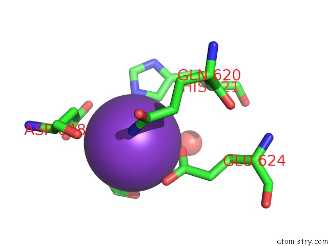
Mono view
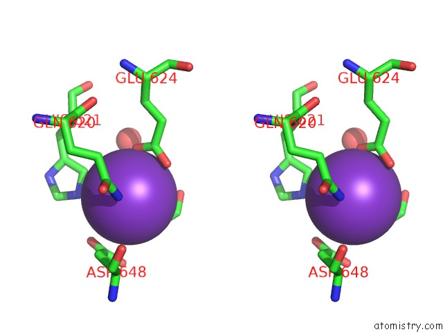
Stereo pair view
|
A full contact list of Potassium with other atoms in the K binding
site number 3 of Crystal Structure of Danio Rerio Histone Deacetylase 6 Catalytic Domain 2 Complexed with to-317 within 5.0Å range:
probe
|
atom
|
residue
|
distance (Å)
|
B
|
Occ
|
A:K910
b:38.8
occ:1.00
|
O1
|
A:FMT904
|
2.3
|
23.7
|
1.0
|
OE1
|
A:GLU624
|
2.8
|
15.2
|
1.0
|
C
|
A:FMT904
|
2.8
|
28.0
|
1.0
|
NE2
|
A:HIS621
|
3.0
|
11.8
|
1.0
|
CD2
|
A:HIS621
|
3.0
|
12.6
|
1.0
|
O2
|
A:FMT904
|
3.1
|
27.6
|
1.0
|
OE2
|
A:GLU624
|
3.1
|
14.5
|
1.0
|
CD
|
A:GLU624
|
3.3
|
15.3
|
1.0
|
NE2
|
A:GLN620
|
3.4
|
9.3
|
1.0
|
OD2
|
A:ASP648
|
3.4
|
10.0
|
1.0
|
CG
|
A:ASP648
|
3.5
|
12.8
|
1.0
|
OD1
|
A:ASP648
|
3.8
|
13.4
|
1.0
|
CB
|
A:ASP648
|
3.9
|
9.9
|
1.0
|
O
|
A:HOH1007
|
4.0
|
24.1
|
1.0
|
CE1
|
A:HIS621
|
4.3
|
9.8
|
1.0
|
CG
|
A:HIS621
|
4.4
|
9.6
|
1.0
|
CD
|
A:GLN620
|
4.5
|
7.8
|
1.0
|
CG
|
A:GLN620
|
4.7
|
6.9
|
1.0
|
CA
|
A:ASP648
|
4.7
|
10.8
|
1.0
|
CG
|
A:GLU624
|
4.9
|
8.6
|
1.0
|
ND1
|
A:HIS621
|
5.0
|
7.4
|
1.0
|
|
Potassium binding site 4 out
of 5 in 7jom
Go back to
Potassium Binding Sites List in 7jom
Potassium binding site 4 out
of 5 in the Crystal Structure of Danio Rerio Histone Deacetylase 6 Catalytic Domain 2 Complexed with to-317
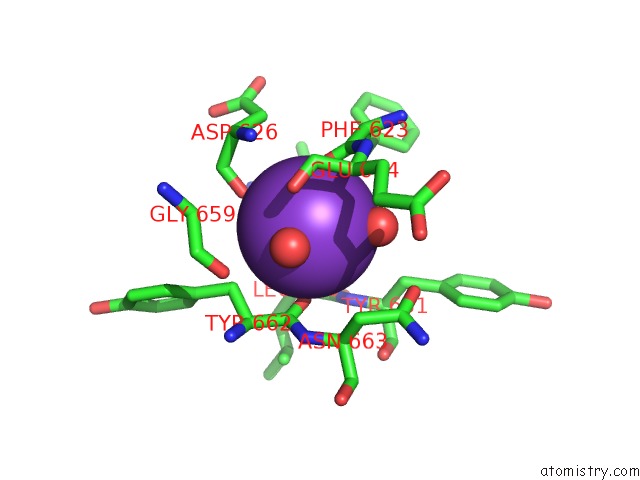
Mono view
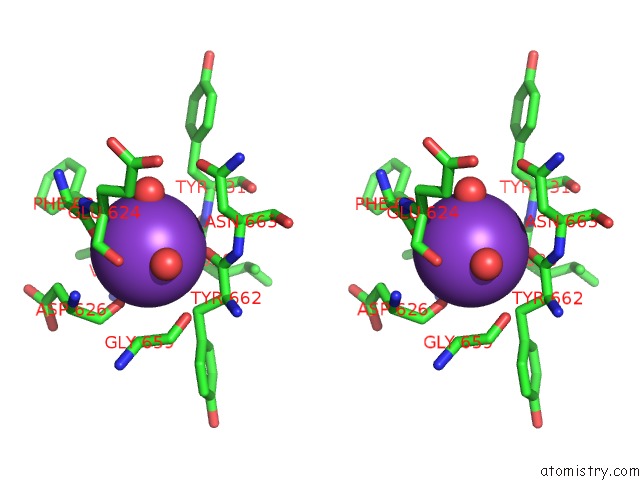
Stereo pair view
|
A full contact list of Potassium with other atoms in the K binding
site number 4 of Crystal Structure of Danio Rerio Histone Deacetylase 6 Catalytic Domain 2 Complexed with to-317 within 5.0Å range:
probe
|
atom
|
residue
|
distance (Å)
|
B
|
Occ
|
B:K805
b:8.4
occ:1.00
|
O
|
B:PHE623
|
2.6
|
6.1
|
1.0
|
O
|
B:VAL629
|
2.7
|
7.8
|
1.0
|
O
|
B:HOH957
|
2.7
|
5.6
|
1.0
|
O
|
B:TYR662
|
2.8
|
6.0
|
1.0
|
O
|
B:HOH1031
|
2.8
|
5.9
|
1.0
|
O
|
B:ASP626
|
2.9
|
10.6
|
1.0
|
C
|
B:PHE623
|
3.6
|
8.1
|
1.0
|
C
|
B:TYR662
|
3.6
|
6.9
|
1.0
|
CB
|
B:TYR662
|
3.7
|
8.7
|
1.0
|
CB
|
B:PHE623
|
3.8
|
5.9
|
1.0
|
C
|
B:VAL629
|
3.9
|
5.2
|
1.0
|
C
|
B:ASP626
|
4.0
|
11.3
|
1.0
|
CA
|
B:TYR662
|
4.3
|
7.5
|
1.0
|
N
|
B:TYR631
|
4.3
|
5.2
|
1.0
|
CA
|
B:PHE623
|
4.3
|
6.8
|
1.0
|
N
|
B:ASN663
|
4.4
|
7.5
|
1.0
|
N
|
B:ASP626
|
4.4
|
8.8
|
1.0
|
N
|
B:GLU624
|
4.5
|
6.8
|
1.0
|
CA
|
B:GLU624
|
4.5
|
5.8
|
1.0
|
CA
|
B:LEU630
|
4.6
|
5.1
|
1.0
|
CA
|
B:ASP626
|
4.6
|
8.2
|
1.0
|
O
|
B:GLU624
|
4.6
|
8.2
|
1.0
|
C
|
B:GLU624
|
4.6
|
8.0
|
1.0
|
CB
|
B:ASP626
|
4.7
|
10.1
|
1.0
|
N
|
B:LEU630
|
4.7
|
5.2
|
1.0
|
CB
|
B:TYR631
|
4.7
|
4.4
|
1.0
|
CA
|
B:ASN663
|
4.8
|
8.3
|
1.0
|
CB
|
B:ASN663
|
4.8
|
5.4
|
1.0
|
C
|
B:LEU630
|
4.9
|
5.7
|
1.0
|
O
|
B:GLY659
|
4.9
|
9.6
|
1.0
|
CA
|
B:VAL629
|
4.9
|
5.2
|
1.0
|
|
Potassium binding site 5 out
of 5 in 7jom
Go back to
Potassium Binding Sites List in 7jom
Potassium binding site 5 out
of 5 in the Crystal Structure of Danio Rerio Histone Deacetylase 6 Catalytic Domain 2 Complexed with to-317
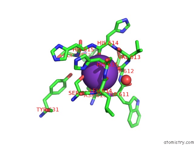
Mono view
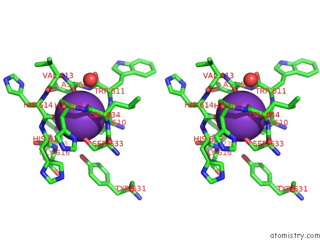
Stereo pair view
|
A full contact list of Potassium with other atoms in the K binding
site number 5 of Crystal Structure of Danio Rerio Histone Deacetylase 6 Catalytic Domain 2 Complexed with to-317 within 5.0Å range:
probe
|
atom
|
residue
|
distance (Å)
|
B
|
Occ
|
B:K806
b:7.4
occ:1.00
|
O
|
B:ASP612
|
2.6
|
5.3
|
1.0
|
O
|
B:LEU634
|
2.7
|
7.9
|
1.0
|
O
|
B:HIS614
|
2.7
|
5.5
|
1.0
|
OG
|
B:SER633
|
2.7
|
7.3
|
1.0
|
OD1
|
B:ASP610
|
2.8
|
7.1
|
1.0
|
O
|
B:ASP610
|
2.9
|
6.0
|
1.0
|
CG
|
B:ASP610
|
3.2
|
6.0
|
1.0
|
C
|
B:ASP610
|
3.5
|
5.9
|
1.0
|
C
|
B:ASP612
|
3.6
|
7.3
|
1.0
|
C
|
B:HIS614
|
3.7
|
5.9
|
1.0
|
C
|
B:LEU634
|
3.7
|
6.1
|
1.0
|
CB
|
B:ASP610
|
3.8
|
6.1
|
1.0
|
N
|
B:ASP612
|
3.8
|
5.0
|
1.0
|
OD2
|
B:ASP610
|
3.9
|
7.4
|
1.0
|
CB
|
B:HIS635
|
4.0
|
6.0
|
1.0
|
N
|
B:LEU634
|
4.0
|
8.1
|
1.0
|
CB
|
B:SER633
|
4.0
|
3.9
|
1.0
|
CA
|
B:ASP612
|
4.1
|
6.3
|
1.0
|
C
|
B:TRP611
|
4.2
|
7.3
|
1.0
|
N
|
B:TRP611
|
4.2
|
5.4
|
1.0
|
CB
|
B:ASP612
|
4.2
|
5.2
|
1.0
|
CA
|
B:ASP610
|
4.3
|
5.9
|
1.0
|
CA
|
B:TRP611
|
4.3
|
5.9
|
1.0
|
CA
|
B:HIS615
|
4.3
|
6.9
|
1.0
|
CA
|
B:SER633
|
4.4
|
5.5
|
1.0
|
N
|
B:HIS615
|
4.4
|
4.8
|
1.0
|
N
|
B:HIS614
|
4.4
|
5.2
|
1.0
|
O
|
B:HOH940
|
4.5
|
8.1
|
1.0
|
ND1
|
B:HIS635
|
4.5
|
7.4
|
1.0
|
CA
|
B:HIS635
|
4.5
|
7.1
|
1.0
|
N
|
B:GLY616
|
4.5
|
6.3
|
1.0
|
CA
|
B:LEU634
|
4.5
|
9.3
|
1.0
|
C
|
B:SER633
|
4.6
|
7.2
|
1.0
|
N
|
B:HIS635
|
4.6
|
7.6
|
1.0
|
C
|
B:VAL613
|
4.6
|
8.0
|
1.0
|
CA
|
B:HIS614
|
4.7
|
5.6
|
1.0
|
CG
|
B:HIS635
|
4.7
|
8.0
|
1.0
|
N
|
B:VAL613
|
4.7
|
3.8
|
1.0
|
OH
|
B:TYR631
|
4.7
|
6.5
|
1.0
|
C
|
B:HIS615
|
4.8
|
6.5
|
1.0
|
CE1
|
B:HIS573
|
4.9
|
6.2
|
1.0
|
O
|
B:TRP611
|
4.9
|
6.4
|
1.0
|
ND1
|
B:HIS573
|
5.0
|
7.4
|
1.0
|
|
Reference:
P.R.Watson,
D.W.Christianson.
Crystal Structure of Danio Rerio Histone Deacetylase 6 Catalytic Domain 2 Complexed with to-317 To Be Published.
Page generated: Mon Aug 12 19:08:13 2024
|